I reach for my phone. The voice on the other end is calm and collected, but I can tell something is wrong. There’s an urgency in the voice, and I know it must be something important. It’s one of many calls I receive each week as the co-owner of Sprig—an innovative pediatric healthcare technology company. Each call is unique. Each call is important. Each call provides me an opportunity to communicate with our customers—sharing advice with them, discussing treatment-plan options, or just letting them know we’re here for them.
This particular call stuck out in my mind because it involved a situation every pediatric dentist dreads—getting a call from the parent whose child’s case was performed under general anesthia (GA) saying something has gone wrong with treatment. Maybe it’s a crown that came out, or a space-maintainer that came loose. Whatever the case, the first thing that goes through our minds is, “What am I going to do? Do I need to put the child back under GA? Are there any other alternatives?” This was the nature of the call I had just received from Dr. Evelyne Vu-Tien, a pediatric dentist in San Diego, California. In the conversation that follows, Dr. Vu-Tien and I discuss a case done under general anesthesia, after which a crown fracture was noticed post-operatively. We explore options regarding what to do when the patient is a young child whose cooperation is limited.
Dr. Fisher: What is your usual restoration choice when restoring lower cuspids, and how have pediatric Zirconia crowns affected that choice?
Dr. Vu-Tien: When I find large interproximal decay in a cuspid or decay involving a cuspid incisal edge, I use Zirconia crowns because they are the most durable restoration out there. Especially in children where bruxing and lateral excursive forces constantly fracture large class III composite restorations, I find that Zirconia crowns are the fastest to place and are the strongest restoration available.
I used to perform large strip-form type crowns. It would take me a long time to polish them down into proper occlusion. Also, they would frequently fracture after a few months or inevitably fail within one to two years after placement. In younger children under 5 years of age, placing a Zirconia crown gives me the peace of mind knowing that it will last them at least the next three to four years. Prior to this case, I had completed about a dozen cases using canine EZCrowns, each with success and without incidence. So, what happened next surprised me.
Dr. Fisher: Tell us about your patient, Julie, and the particulars of her case.
Dr. Vu-Tien: I saw Julie in November 2015, when she presented for her first initial exam at age 5. We discovered she had 12 cavities. She was extremely apprehensive and anxious, and it was difficult to coax her to open her mouth for a visual examination. Due to the severity and amount of decay, as well as her level of apprehension, her mother and I discussed monitored-anesthesia care for her treatment.
We ended up placing six crowns and six posterior interproximal restorations. We placed EZCrowns on teeth E, F, M, and R with no pulpotomies required. I decided to substitute upper canine crowns for the lowers, using a smaller size (Size 1 of C and H). I also used Fuji Cem for cementation instead of the recommended Ketac Cem. I did not hear any “snap” or “breaking” sounds upon seating the crowns, nor did I notice any fracture lines after the treatment was completed.
Dr. Fisher: In Julie’s case, when did you first notice the fracture in the crown, and what went through your head in the moments that followed?
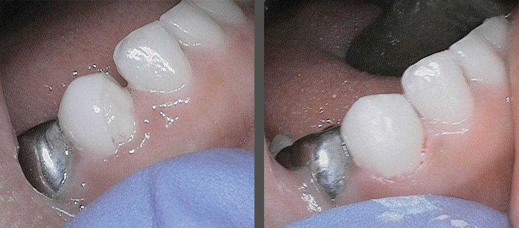
Dr. Vu-Tien: I received an evening call from Julie’s mother one week after the surgery, alerting me that a piece of the “tooth” had broken on the lower right area of the crown. I did not believe her at first. I had never had a canine crown fracture before. I immediately wondered if it was a piece of excess cement that I had failed to remove, or if it was a chip on a neighboring tooth. I tried to recall if I heard any “snap” or felt any “break” during cementation, but I could recall nothing. I asked her to text me a photo of the tooth right away. Sure enough, a diagonal piece of the crown was missing. Julie’s mother said her daughter had been eating soft foods at the time and handed mom the chipped piece.
I was worried how I would be able to restore the fragment chair side and wanted to look up what materials bond to Zirconia. Luckily, the cement under the crown was still present, and Julie was not having any pain or sensitivity. I did not want Julie to undergo anesthesia again to repair just one tooth, so I was trying to brainstorm ideas on how to “patch it” until she could tolerate a chair-side removal of the crown or until the tooth would exfoliate naturally in two to three years.
Dr. Fisher: What are your thoughts on why the crown might have fractured?
Dr. Vu-Tien: It was puzzling, because I had placed two lower canine EZCrowns, only one of which chipped. At the time I performed the case, I did not have the SL cuspid sizing, so I placed regular canine crowns instead. The other canine crown (M) did not fracture. I suspect that I may have twisted the lower right crown (R) upon cementation, which led to the failure and fracture of the crown. The regular-sized cuspid Zirconia crowns are more ideal for placing on maxillary primary cuspids, while the SL-sized cuspids— being somewhat narrower—are more suitable for use on lower canines. I also wondered if by using the Fuji Cem, which was less viscous than Ketac Cem—if it somehow contributed to decreased retention of the crown, resulting in failure where the chip occurred.
Dr. Fisher: When you called me, you had a good idea of what you wanted to do to fix the problem. What was your idea?
Dr. Vu-Tien: When I called you, I wanted to review my options with you and discuss what experience you may have had with this type of fracture. Because of Julie’s anxiety level, the idea of removing the crown with a high-speed hand piece, requiring water, suction and re-cementation of a new crown was something highly unfeasible given her level of anxiety. Julie barely spoke any English, and it was challenging trying to communicate with her. Typical “tell- show-do” methods and voice control had not proved effective. Rather, I opted to repair the tooth with composite to buy us some time until she was older and could better tolerate more definitive treatment. I did not want Julie to have to undergo anesthesia an additional time for repair of a crown fracture that was neither symptomatic nor carious.
Dr. Fisher: What were the procedures you used to fix Julie’s broken crown?
Dr. Vu-Tien: When Julie returned to the office, she was still very anxious. It was difficult to place the nitrous mask on her. Upon inspection of tooth R, we discovered the remaining unchipped area of the EZCrown was intact and well cemented. Since cement still covered the exposed portion of tooth, I opted to bond composite to this exposed area.
I first cleaned the area with chlorehexidine and used a size-2 round bur to roughen the cement and clear out any debris. I used a mouth prop and cotton roll isolation in lieu of a rubber dam because of Julie’s behavioral issues. After placing a sectional matrix, I applied 35% phosphoric etchant and adper bond (Scotchbond) to the cleaned surface. I applied A1 flowable composite (Shofu) to the interface between the crown and tooth and followed up with Filtek Supreme color B1 over the open area. I then polished using football and flame-shaped carbide burs to contour the composite. Finally, I polished with a Shofu white point to achieve smoothness and shine. I advised Julie’s mom that the composite restoration possibly could chip and stain and may need revisions in the future, but at this time it was a viable option we had chosen instead of replacing the entire crown while Julie is still young and apprehensive. Julie’s mom was very happy with the result.
Dr. Fisher: were there any challenges with the “zirconia repair,” and what did you learn from this case?
Dr. Vu-Tien: Julie’s mom was very happy with the results and with the fact that we could achieve them without additional monitored anesthesia care. That joy was short-lived, however, because one month later, I got a call from Julie’s mom telling how the crown had fractured again. This situation allowed me an opportunity to reflect yet again on how I might have repaired the tooth better.
I requested Julie to return, and we removed all the old cement to expose the dentin. I then followed the steps outlined above to ensure a good bond. This time, I took the tooth out of occlusion by 1–2 mm and rounded the edge of the canine with a new diamond and lots of water to prevent heating the tooth. Julie’s mom was pleased with the second repair.
I contacted mom again one month later to check on Julie. I learned she was doing great, experiencing no issues and with the filling still intact.
Dr. Fisher: having repaired a fractured zirconia crown now in your own practice, what would you advise your colleagues to do if they were to encounter a similar issue in their practice?
Dr. Vu-Tien: First off, I would pause and reflect on the steps taken to complete the crown. Could the error have been caused by one of the following factors? 1) inadequate tooth preparation, 2) crown choice (SL vs. regular sizing), 3) cementation process, or 4) type of cement. Answering these questions allows you to critically evaluate each of the steps taken. You can then more accurately deduce how you might have performed the procedure better. This process also allows you to review how you could have given more careful attention to the materials used or steps taken, possibly ensuring a better outcome.
I am thankful for a humbling experience such as this because I can learn and grow from my mistakes. I am so appreciative that you, Dr. Jeff, were always available to speak with me and offer support, guidance, and mentorship throughout the process. This is the only company I know of that is owned and managed by dentists with a passion for educating their dentist customers and ensuring that their product improves a patient’s overall health and well-being.
Often in the practice of dentistry we are presented with situations that are less than ideal. It’s just the nature of the profession, and we as practitioners need to be able to use critical thinking to solve these challenging situations. The above-mentioned case is just one example of any number of situations—many of which you might decide to resolve differently. That’s the beauty of private dental practice. Our education has equipped us with tools to use in the treatment of our patients. It is up to us, as individual dentists, to use that knowledge to offer the best treatment we can to those seeking our help.
As the horizon of pediatric dentistry continues to expand, we will be faced with more and more situations requiring us to exercise our critical-thinking skills. I want to encourage all of us to think outside the box. Think with a critical mind. Use the “tools” you must evaluate every new product and technique that you encounter. Learn and decide for yourself how you will respond to the most important question you face every day in practice: “What is the best for my patient?”


